


Best Neurologist in Dubai | Neurology doctor | Pediatric Neurologist Dubai

🧠 Neurology Doctor in Dubai – Discover Advanced Brain & Nerve Care at KindCare
According to the well-known expression, “All diseases are from nerves,” the demand for expert neurological care has never been greater. At KindCare Medical Center, our Neurology Department provides specialized care for disorders of the Central Nervous System (brain and spinal cord) and Peripheral Nervous System (nerves and ganglia). If you’re searching for a reliable neurology doctor or the best neurologist in Dubai, you’ve come to the right place.
🌟 Why Choose KindCare for Neurology in Dubai?
KindCare stands out as one of the most trusted destinations for people looking for a neurologist in Dubai. We offer a unique approach by combining:
Integrative Medicine 🧬
Digital Diagnostics 🧪
Evidence-Based and Alternative Treatments 🌿
This combination, often referred to as the “Medicine of the Future,” ensures that each patient receives personalized, effective, and forward-thinking neurological care.
🧾 Conditions We Treat – Comprehensive Neurological Care
Our expert neurology doctors in Dubai specialize in the prevention, diagnosis, and treatment of various neurological conditions, including:
🤕 Chronic headaches and migraines
⚡ Epilepsy and seizure disorders
🧠 Stroke and post-stroke care
🧍 Multiple sclerosis
🕰️ Parkinson’s disease
🔥 Neuropathy and nerve pain
😴 Sleep disorders
🧓 Alzheimer’s and memory issues
💃 Movement disorders
🧒 Pediatric neurological disorders
Whether you’re searching for the top 10 neurologists in Dubai or simply need a neurology doctor near me, KindCare is here to help.
🔍 Symptoms That Require a Neurology Doctor in Dubai
Neurological issues can manifest in many subtle and serious ways. You should consult a neurology doctor near me if you or a loved one experiences:
Persistent headaches or migraines
Dizziness or balance issues
Memory loss or cognitive decline
Unexplained numbness or tingling
Seizures or convulsions
Sleep disorders
Chronic fatigue
Neck or back pain radiating to limbs
At KindCare, our goal is not just to treat symptoms, but to find and eliminate the root causes.
👨⚕️ Meet Dr. Timur – One of the Top 10 Neurologists in Dubai
Dr. Timur, a board-certified neurology doctor at KindCare, is widely recognized as one of the top 10 neurologists in Dubai. He combines deep clinical knowledge with a compassionate approach to patient care. His expertise includes:
Stroke and post-stroke rehabilitation
Epilepsy
Multiple Sclerosis
Parkinson’s Disease
Alzheimer’s and other cognitive disorders
Neuropathy and nerve compression syndromes
Patients looking for a neurologist near me or the best neurologist Dubai consistently choose Dr. Timur for his results-driven, integrative care.
🧒 Pediatric Neurologist in Dubai – Gentle, Specialized Care for Children
At KindCare, we understand that neurological issues in children require a different approach. Our dedicated pediatric neurologist Dubai team provides comprehensive evaluation and treatment for conditions such as:
ADHD and behavioral disorders
Epilepsy and seizures
Developmental delays
Autism Spectrum Disorders
Headaches and migraines in children
If you’re seeking the best pediatric neurologist Dubai has to offer, our specialists are equipped with the knowledge, patience, and tools to guide your child’s recovery and development.
🧪 Advanced Neurological Diagnostics at KindCare Dubai
Our clinic utilizes modern tools and technologies to ensure accurate diagnoses:
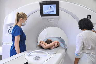
MRI & CT Scans
EEG (Electroencephalography)
EMG (Electromyography)
Nerve Conduction Studies
Balance Testing & Vestibular Evaluation
Digital Cognitive Assessment
These allow your neurology doctor to pinpoint the exact origin of your symptoms and plan precise interventions.
🎯 Personalized Treatment Plans – Tailored to You | Neurologist Near Me
We believe that every brain is unique 🧠—which is why our neurology doctors create personalized care plans. Whether you’re struggling with chronic migraines or recovering from a stroke, our team ensures you’re receiving care that fits your needs and lifestyle.
🏥 Why Dubai Residents Trust KindCare Neurology Clinic | Neurologists Dubai
Here’s why patients consistently choose KindCare:
🌍 Internationally trained neurologists
🔁 Integrative and conventional medicine combined
👨👩👧👦 Family-friendly environment
📞 Easy appointment booking
🥇 Rated among the best neurologists in Dubai
Looking for a neurologist near me who truly cares? You’ve found one.
🌍 Conveniently Located – Find a Neurologist Near Me in Dubai
Whether you’re living in Jumeirah, Business Bay, Al Barsha, or Downtown Dubai, KindCare’s central location makes it easy to reach a neurologist near me without the hassle. We’re strategically located with easy access to parking, public transport, and in-house diagnostics—all under one roof.
📈 The KindCare Promise: Excellence in Neurology Dubai
When it comes to your brain and nervous system, trust only the best. KindCare’s Neurology Department:
Helps hundreds of patients every year
Maintains high patient satisfaction scores
Offers multilingual staff for easy communication
Is recognized by leading insurance providers in the UAE
Patients in search of the best neurologist in Dubai rely on KindCare for expert care, genuine empathy, and lasting results.
📆 Book Your Appointment with a Neurology Doctor Today!
Don’t wait until your symptoms get worse. Early diagnosis saves lives and improves quality of life. Whether you need a neurologist in Dubai, a best pediatric neurologist in Dubai, or just a second opinion, we’re here for you.
➡️ Call or book online today to meet your trusted neurology doctor in Dubai!
✨ KindCare Medical Center – Leading Neurology Clinic in Dubai
Advanced treatment, modern tools, and compassionate care under one roof.
Neurology Diagnostic Tests for Children & Adults in Dubai


Effective Help for Children & Adults
Pharmacotherapy is one of the main methods in the treatment of many neurological diseases. To effectively restore and treat the function of the Central and Peripheral Nervous System (CNS and PNS), and to eliminate the cause of the disorder, the correct combination of medications is needed.
Botulinum toxin therapy is a method of treating Neurological Disorders by injecting botulinum toxin type A. After injecting botulinum toxin into a muscle, the transmission of nerve impulses to the muscle is "switched off" and the muscle is paralyzed. This method relieves spasticity, spasms, pain syndromes, treats spastic torticollis, migraine, alleviates cerebral palsy symptoms, autonomic disorders, tics, tremors and others.
Therapeutic blockade in neurology is recognized as the most effective method. With the help of a blockade, not only the pain syndrome is relieved, it also has a therapeutic effect. Often, after the first procedure, patients forget about their diseases. It is effective for acute and chronic pain syndromes in the cervical, thoracic, lumbar spine, osteochondrosis, intervertebral bulges, disc protrusions, knee, shoulder joints, etc., as well as for the treatment of acute and chronic pain syndromes in the cervical, thoracic, lumbar spine.
The use of ozone therapy Dubai (ozone-oxygen mixture) in neurology is a highly effective non-medication method of prevention and treatment of Cerebrovascular Pathology, Cerebral Atherosclerosis, Cerebral Ischemia, Tinnitus, Fatigue, Memory Impairment, Autonomic Dystonia, Toxic (Alcoholic) Polyneuropathy, Parkinson's Disease, Alzheimer's Disease, Inflammatory Diseases of the Brain, Multiple Sclerosis, Sleep Disturbance, Mood Improvement (Natural Antidepressant), etc.
Complex Vitamin Drips, including more than 30 ingredients (exclusive to us), individually selected for your Neurological Concerns with 100% absorption in the body. The protocols developed for this therapy and drug combinations are unique and patented by us. The earliest symptoms of vitamin deficiencies occur in the Nervous System, such as Mental Changes, Irritability, Fearfulness, Hallucinations, Loss of Concentration and Attention, Memory Loss, Impaired Mental Ability, Depression, Sensitivity, Paresthesia, Neuropathic Pain, Muscle Weakness, Paralysis, Muscle Atrophy, etc.
Antioxidants are substances that block or inhibit oxidation, as a manifestation of the negative and destructive effect on the brain and nervous system, by free radicals. Antioxidants are effective in protecting against existing free radicals and inhibiting their formation. Deficiency of even one of the Amino Acids can worsen the condition of the Nervous System and lead to serious neurological disorders. Amino acids are better taken by injection to guarantee their absorption in the orgasm.
Amino acids will help to improve recovery from injuries, speed up and improve the recovery processes of brain activity. Amino acids increase mental performance and relieve excessive psycho-emotional stress.
Minerals are essential vital components of body tissues. At the same time, the body cannot synthesize them independently. In addition to the basic minerals - carbon, hydrogen, oxygen and nitrogen, which a person gets without additional efforts from natural sources, there are also potassium, chloride, sodium, calcium, phosphorus, magnesium, iron, zinc, etc., the deficiency of which has a detrimental effect on the function of the nervous system, brain and physical activity of the body, such as loss of memory, impaired concentration, dizziness, muscle spasms, intellectual and developmental dalay, etc.
Heavy metals are elements that even at low concentrations can be toxic and very dangerous to the human body and, once in the body, their compounds have a toxic effect, causing a number of neurological diseases. The most alarming representatives of this group are arsenic, cadmium, lead and mercury. Only in our clinic, we offer the only way to eliminate heavy metals - Chelation Therapy, which is a chemical process where a synthetic solution of EDTA (ethylenediaminetetraacetic acid) is introduced into the bloodstream via injection to remove heavy metals/minerals from the body. When the EDTA solution is injected into a vein, it "binds" heavy metals and minerals (such as lead, mercury, copper, iron, arsenic, aluminum and calcium) and removes them, helping to treat Chronic Fatigue, Autoimmune Diseases, including Lyme disease, neurological disorders, decreased brain activity, poor concentration, learning difficulties and poor memory, depression, anxiety, dementia, insomnia and more.
In our Clinic, different physicians are invited to make a complete diagnosis of neurological diseases. Comprehensive assessment of the patient's health condition and examination of several specialists helps to choose a comprehensive treatment consisting of a minimum set of procedures, but solving several problems of the patient at the same time. Specialists in a joint discussion establish the treatment goal, objectives, time of their solution, based on the patient's needs. When all problem areas are identified and goals are set, the treatment team forms a treatment program. This approach makes it possible to optimize the diagnostic and treatment processes, ensuring the best result in the patient's recovery.
Neurofeedback "Fitness for the Brain" The brain remains the most mysterious organ of the human body. In the process of studying how the brain works, it was discovered that it can be specifically influenced and positive changes can be achieved through EEG-based biofeedback called "Neurofeedback", in which the electrical activity of the brain is recorded with the help of QEEG (Quantitative Electroencephalogram) in real time and its results are displayed through various sounds and images in the form of a video game on a computer monitor, in the process of which training of the nervous system takes place, which can be used to correct ADHD (Cognitive Behavioral Disorder), Epileptic syndromes, CNS dysfunctions after strokes, traumas of the brain, as well as for Migraine, Depression, Anxiety, Tics, Autism, as well as Preventing Alzheimer's Disease and others.
Biofeedback is a method that helps to achieve positive results in the treatment of neurological diseases. With the help of special equipment and computer technologies that receive and analyze brain signals, the patient learns to control his/her body and thus regulate his/her own healing process. The aim of the method is to show the patient that he is able to control his body and mind in any situation and to give him the skills to manage his condition. This method of treatment is used for the treatment and prevention of such diseases as Headache, Migraine, Hyperactivity, Depression, Fears and Phobias, Psychosomatic Disorder, Bipolar Disorder, Panic Attacks, Stress, Sleep Disorder, Chronic Fatigue, Stuttering, Speech Disorders, Obsessive-Compulsive Disorder, Autism, Epilepsy and others.
Microcurrent Reflexotherapy is a popular rehabilitation method of treatment that demonstrates high efficiency in the process of restoring the condition of children diagnosed with developmental delays due to neuropsychiatric diseases. Microcurrent Reflexotherapy uses ultra-low electrical signals that are applied to various biologically active points to restore the patient's own normal brain and spinal cord function. Due to preliminary examination and individual approach to each patient, Microcurrent Reflexology helps in treatment of children with such neurological diseases as Speech delay, Developmental delay, Autism, Cerebral palsy, learning problems, increased intracranial pressure, hearing loss, Attention Deficit and Hyperactivity Disorder and others.
Our Nervous System is securely protected and hidden from external influences, which becomes an obstacle for effective and painless treatment. Therefore, in the treatment of neurological disorders is well-proven percutaneous electroneurostimulation (PENS), which consists in the action of a weak electric current on the painful areas, helping to stimulate nerves, which, in turn, improves well-being, increases blood flow in the damaged organ and reduces pain. This method of physiotherapy helps in the treatment of Chronic Muscle and Joint Pain, Bursitis (inflammation of the synovial sac), Arthritis and Osteoarthritis, Tendinitis (tendon damage), Oncological Diseases to reduce pain, etc.
Transcranial Magnetic Stimulation (TMS) is a modern non-invasive technique that allows stimulating nerve cells in the affected areas of the brain, which leads to their activation and inclusion in the process of providing speech and higher mental functions of the patient. This procedure helps to restore nerve connections in the cerebral cortex and enables non-invasive targeted stimulation of cortical structures. Depending on the mode chosen by the specialist, the effect on the central nervous system can be either excitatory or inhibitory. Regardless of the type of influence, intercellular interaction and all types of metabolism are improved in the tissues of the cerebral cortex, and blood microcirculation is normalized.
This procedure improves cognitive function in children and adults with a wide range of central nervous system disorders such as Alalia, Aphasia, Dysarthria, Speech delay, Developmental delay, Autism Spectrum Disorders (ASD), mental retardation, genetic syndromes, stuttering, epilepsy, consequences of brain injuries, strokes, amnesia, etc. The use of TMS is not recommended for patients who have an electronic pacemaker, in the presence of large aneurysms of cerebral vessels. It is also not recommended for pregnant women and patients taking large doses of anticonvulsants.
Bioresonance therapy is a therapy with the body's own healthy biological electromagnetic vibrations after their special processing and their correct selection, which is proven by scientists Dr. Franz Morel and Erich Rasche. Healthy biological electromagnetic vibrations bring the human body and individual organs and systems into harmony, as well as train the nervous system to emit the correct and desired neuroimpulses. The device registers real-time parameters of the organism and individual organs, converts pathological (bad) physiological vibrations into good ones and sends them back to the organism to create resonance (Resonance is a sharp increase in the amplitude of vibrations when frequencies coincide). Thus, the patient and the device form a closed loop of regulation, where specially processed and correctly selected frequencies of electromagnetic oscillations return to the patient again and again with healthy physiological oscillations. This therapy is widely used in the treatment of diseases of the Central Nervous System and Sensory Organs, diseases of the Autonomic Nervous System, such as cerebral palsy, speech and development delay, ADHD, Autism spectrum disorders and others.
Micropolarization is a non-invasive method of treatment that allows changing the functional state of various parts of the Central Nervous System under the action of direct current of up to 1 mA (microcurrents). The method is most recognized in child neurology in the treatment of organic lesions of the CNS, including Cerebral Palsy, delays in neuropsychiatric development and learning problems, speech development disorders in children, psychoemotional, neurotic, Psychosomatic Disorders (Depressive States, Tension Headaches, Hyperactivity, Psychogenic Enuresis and/or Encopresis, Aggression, Fears, Tics), Visual and Auditory dysfunctions, treatment of the consequences of traumatic brain injury and Neuroinfectious Diseases of the brain.
Therapy according to the method of R. Voll - stimulation of biologically active points corresponding to certain organs, using low-frequency current. The mechanism of action of this current has a regulatory nature with a predominant influence on the structures of the nervous system, which is designed to control physical, chemical and electrical processes in the intracellular and extracellular space. This method of therapy helps in Activation of Speech Areas of the Brain (for Speech Development), Activation of Motor Areas of the Brain (for Development of Motor Skills), Learning Areas of the Brain (for Development of Counting, Writing and Reading Skills), Stabilization of Stem Structures (to reduce agitation), Activation of Cerebellum (to improve Movement Coordination), Stabilization of Muscle Tone (for cerebral palsy), Stabilization of Intracranial Pressure, etc.
Treatment of parasitic infections with the help of bioresonance therapy (BRT) is manipulation with electromagnetic vibrations, due to which the body enters into resonance (Resonance is a sharp increase in the amplitude of vibrations at the matching of frequencies), thereby leading the parasite to destruction, or forcing it to leave the body. Approximately 90% of chronic diseases are caused by parasites, as well as toxins that have somehow gotten into the human orgasm, which can lead to headaches, migraines, depressive states, irritability, chronic fatigue, sleep disorders, dizziness, stress, neurosis, tics and others.
Acupuncture is a non-pharmacological method of Prevention, Complex Treatment and Rehabilitation of a number of diseases of the Nervous System, recognized by the Official Medicine. The positive effect of stimulation of biologically active points on the body has been proven by centuries-old practice for more than 4,000 years. A needle inserted into a biologically active point creates nerve impulses that spread through the nearby spinal segment or metameres and activate the subcortical centers of the brain. This effect activates the work of organs and systems, stimulates natural recovery processes, triggers the body's immune response. Acupuncture is effective in neurology for Enuresis, Tics, Depression, Neurosis, Panic Attacks, Stuttering, Central Nervous System damage, Motor, Sensory and Speech Disorders, Cardiovascular Disorders and Brain and Spinal Cord Injuries. The analgesic efficacy of acupuncture has been proven for all types of pain.
It is a treatment method developed by a South Korean scientist, Prof. Park Jae Woo. Su - in translation from Korean - means hand, Jok - foot, that is, it is a treatment using hands and feet, where hands and feet are similar to the body in structure. They contain bioenergetic points of correspondence of all organs and systems, affecting which with the help of specially designed needles, causes increased blood circulation in a given organ and the production of biologically active substances, which thus helps in the healing of this organ and the body as a whole. In neurology, this therapy is used for various manifestations of Spine disorders, disc bulges, sciatica, neuritis, neuropathic pain, migraine, chronic and post-traumatic headache, dizziness, sleep disturbance, stress, multiple sclerosis, migraine, etc.
It is a treatment method developed by a South Korean scientist, Prof. Park Jae Woo. Su - in translation from Korean - means hand, Jok - foot, that is, it is a treatment using hands and feet, where hands and feet are similar to the body in structure. They contain bioenergetic points of correspondence of all organs and systems, affecting which with the help of specially designed needles, causes increased blood circulation in a given organ and the production of biologically active substances, which thus helps in the healing of this organ and the body as a whole. In neurology, this therapy is used for various manifestations of Spine disorders, disc bulges, sciatica, neuritis, neuropathic pain, migraine, chronic and post-traumatic headache, dizziness, sleep disturbance, stress, multiple sclerosis, migraine, etc.
Physical factors have a special influence on the state of physiological systems of the organism. Today one of the safest methods of treatment of pathologies of the Peripheral and Central Nervous System is Physiotherapy - stimulation, conducted with the help of electric current or magnetic fields, the effect of which is aimed at the treatment of a number of pathologies, such as neuropathy, neuralgia, atherosclerotic encephalopathy, cerebral circulatory disorders, cerebral infarction of ischemic and hemorrhagic types, neurological manifestations of degenerative and dystrophic diseases of the spine, and others.
In the treatment of many diseases, no medicine can replace movement - this has been known since ancient times. Therapeutic Exercises for diseases of the nervous system takes a very important place in the recovery processes and complex treatment. Therefore, the use of therapeutic exercises increases the mobility of nervous processes, promotes the development of new neuron connections and the formation of optimal motor stereotypes, which, in turn, help in the rehabilitation of neurological diseases such as stroke, brain injury, cerebral palsy, cerebral palsy, ADHD, neurosis, depression and other functional disorders.
The nervous system of the body plays an important role in maintaining homeostasis and health. Chiropractic is one of the methods of Unconventional Medicine that is effectively used in Neurology. By manipulating the bones and associated muscles and joints, especially in the spine, chiropractic practitioners correct misalignments, thereby improving the functioning of the neuromuscular system and attempting to restore homeostasis to treat conditions such as Migraines and Headaches, Dizziness, Dystonia and other Nervous System problems.
Therapeutic Massage has been known since ancient times and is considered one of the most effective therapeutic treatments. In the treatment of neurological diseases, Therapeutic Massage helps to relieve nervous tension and stabilize the emotional state, gives the patient strength to fight the disease, helps him to recover faster and return to normal life. Thus, Therapeutic Massage is used in Rehabilitation after injuries, surgeries and prosthetics, rehabilitation after stroke and restoration of mobility, Scoliosis, cerebral palsy, ADHD, flat feet, "clubfoot", Headaches and Migraines, Speech Disorder, Neuroses and Tics, Muscular Insufficiency, for relaxation of the Nervous System.
Dry Immersion has a beneficial effect on all physiological processes, naturally improving the homeostasis of the whole organism, providing Relaxing, Spasmolytic, Analgesic, Diuretic and Immunomodulating effects. During immersion children with hyperexcitability, anxiety, neurosis not only quickly calm down, but also improve their behavior and external signs of the disease by normalizing blood flow and intracranial pressure. Immersion quickly releases tension and relaxes not only muscles and internal organs, but also relieves spasm of blood vessels, nerves, bronchi, which gives relief to children with spastic cerebral palsy, which also determines the effectiveness of the course of their rehabilitation. In just 40 minutes, the body is fully restored as during 8 hours of healthy sleep, helping the nervous system to maximize relaxation and effectively affect the rehabilitation of various neurological diseases such as cerebral palsy, hyperactivity syndrome (ADHD), anxiety, neuroses, sleep disorders, stress, etc.
Ayurveda as medicine is over 5,000 years old. Healers of all times have understood the importance of detoxification. The body's detoxification system needs a periodic kick-start for improved elimination of toxins that damage our Nervous System. Ayurveda for Neurological Diseases are natural therapeutic procedures and preparations for prevention, treatment and restoration of proper functioning of the Nervous System, getting rid of Stress, Neurosis, Depression, Insomnia, Memory Loss, Radiculitis, Osteochondrosis, Chronic Fatigue Syndrome, Strokes, Paralysis, Injuries, etc., as well as for the prevention, treatment and restoration of proper functioning of the Nervous System.
Treatment of diseases of the Central Nervous System with herbs, fruits, and flowers is widely known in folk medicine, with the help of which you can get rid of a number of problems in the Nervous System, thereby preventing serious consequences. Herbal medicine is used primarily for Migraine, Consequences of Stroke, Epilepsy, Depressive States, Sleep Disorders, Psychosis, Stress, Panic Attacks, Pain and a number of other Neurological Pathologies. In each specific case, only after the Doctor has identified the cause of the disease (etiology), but also the logic of its development (pathogenesis), in a laboratory with more than 1000 herbs, a decoction from the herbal collection will be prepared for you. The composition, duration and sequence of administration depend on the severity of your disease.
Treatment and rehabilitation of Neurological Diseases includes comprehensive treatment programs with Psychotherapy by a Clinical Psychologist. Shifting the patient's attention from the past to the future, from losses to available opportunities and prospects. Increasing the patient's motivation to recover or adapt to life with chronic or hereditary neurological diseases and how to live with them and cope effectively can help Psychologist with Behavioral Therapy, as well as teach you new ways to solve troubling problems and help in setting real life goals and their implementation.
The environment of large cities often contributes to low levels of oxygen in the air. The lack of oxygen has a negative impact on health, especially on the respiratory and circulatory system. In this regard, the body is in a state of hypoxia. Lack of oxygen negatively affects the Nervous System, impairing the quality of life. Therefore, it is very important to resort to oxygenation of the body through the respiratory system through "Oxygen Therapy", which will help in oxygenation of the body and in the treatment of neurological diseases such as Autism, Speech Delay, Alalia, Hyperactivity, Brain Dysfunction, Depression, Headaches, Cerebral Palsy and others.
Bioacoustic Correction method is a sensory stimulation of the brain with sounds generated on the basis of real-time EEG to activate self-regulation mechanisms.
This method is widely used in pediatric neurology to restore brain functions in patients with neuropsychiatric disorders of various etiologies, such as ADHD, Speech and Development Delay, Alalia, Dysarthria, Stuttering, Autism, Neurosis, Tics, Traumatic Brain Injury, Dysgraphia, Dyscalculia, Dyslexia, Adjustment disorders in preschool and school groups, etc.


